Understanding health at the cellular level
Biochemists and molecular biologists in the College of Natural Sciences are fascinated by the microscopic, tiny building blocks of life: cells. Cells are immensely complex, and studying how they affect health at a single-cell level enables scientists to understand how they come together to create living systems. A deeper understanding of our cells is critical to advancing human health.
The mystery of RNA localization
Erin Osborne Nishimura, professor in the Department of Biochemistry and Molecular Biology, has always been interested in genes, and how gene expression leads to the amazing, diverse animal bodies present on Earth.
Nishimura’s research is at the forefront of understanding gene expression at the single-cell level.
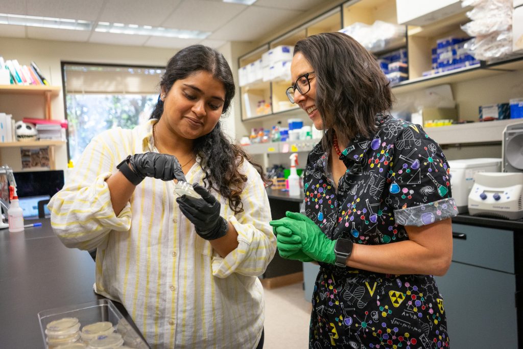
“My lab conducted one of the first studies that produced a map of gene expression at single-cell resolution during early stages of development using Caenorhabditis elegans, a nematode round worm, as our model system,” said Nishimura. “In that study, we discovered something interesting – RNA congregates at different places in the cell. They were not just floating around inside cytoplasm like you would see in a textbook, but they were localizing at weird little corners of the cell. We found lots of different patterns.”
These patterns were found again and again in other research projects; yet, molecular biologists are just beginning to understand the purpose of this behavior.
We discovered something interesting – RNA congregates at different places in the cell.
Erin Osborne Nishimura, professor,
Department of Biochemistry and Molecular Biology
“Sometimes, it is for very important reasons; the location in the cell helps regulate their translational activity or gene expression. But in other cases, we find that it is for no reason, and we are still trying to puzzle through that,” said Nishimura.
One significant advancement in this research is the finding that human neuron cells contain similar patterns to what Nishimura observed in C. elegans.
While we don’t yet know where this discovery could lead, this information could have implications for neuron disorders, such as amyotrophic lateral sclerosis.
“Neurons have been known for a long time to concentrate their RNAs in a certain place within the neuron. Many neuronal diseases are associated with a failure to localize RNAs to different places. This has been a mystery for a long time. Now that we have these more tractable examples of RNA localization, we are hopeful that the information that we learn about how RNAs move around and congregate in C. elegans could lend insight into how the same thing is happening in neuronal cells,” said Nishimura.
Understanding specialized cell functions
Cells need to function in precise and specialized ways to maintain healthy organisms. They do this by using myriad processes that are constantly taking place inside our bodies.
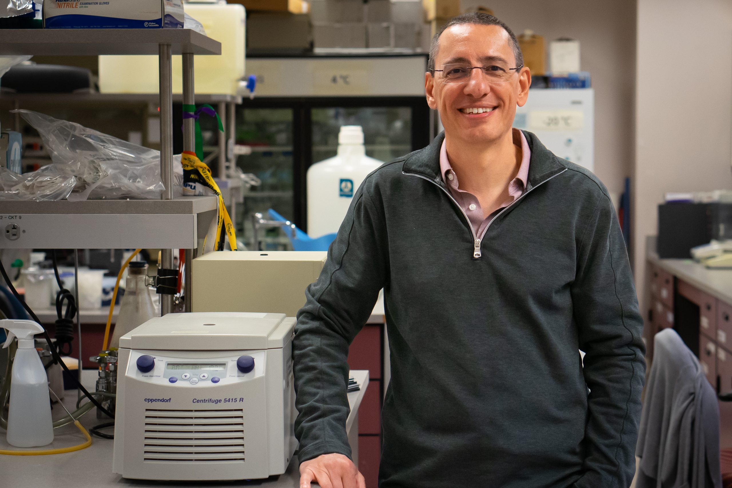
These processes, and what makes them function or malfunction, are of particular interest to Santiago Di Pietro, research associate dean in the College of Natural Sciences. Di Pietro studies a range of cell functions including endocytosis – how cells intake external materials – and specialized cell compartments such as those found in platelet and skin cells.
“In all cases, we are trying to understand at a molecular level how these processes are accomplished,” said Di Pietro.
One example of Di Pietro’s work is with the platelet cell – platelets are anucleate cells that cause blood to clot when there is an injury so that animals do not lose too much blood. To do this they must be in perfect balance; too little activity and a person bleeds too much, too much activity and the platelets can congregate and cause thrombosis, the formation of clots in blood vessels that can lead to adverse health effects such as a heart attack.
A recent finding from Di Pietro’s lab uncovered how proteins affect platelet function inside specialized cell compartments called granules.
“We found that mutations cause proteins that normally accumulate in granules to be incorrectly transported and degraded, resulting in platelets that are missing hundreds of proteins that are needed for normal function,” said Di Pietro.
“These data provide a cellular and molecular understanding of poorly studied, platelet-based bleeding disorders. With the knowledge that we are generating, we can hopefully develop a more targeted way to diminish platelet activity, so you don’t have thrombosis, but you preserve the normal functions of the platelet.”
In addition to platelets, Di Pietro studies skin and eye cells that contain specialized melanin compartments that are critical for normal eye development and defending skin against UV radiation. Di Pietro is also interested in the process of endocytosis, which is necessary in healthy cells but also opens a pathway for infectious agents, such as viruses, to enter cells.
Decoding neuronal communication
How does the human brain work and analyze information?
It turns out that a major cell type in our brain called neurons can process various signals and then spread that message among each other. This form of neuronal communication happens through a specialized microscopic compartment called synapses, which leads to all our brain functions from breathing to talking to moving. Soham Chanda, an assistant professor in the Department of Biochemistry and Molecular Biology, is currently investigating how these synaptic structures form and function.
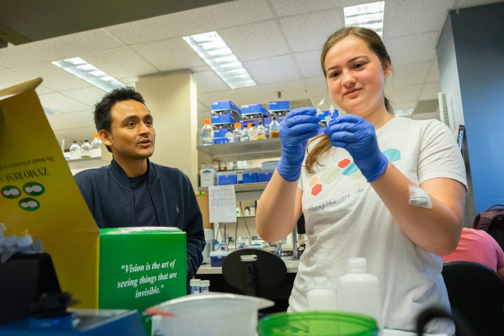
“My lab is focused on molecular neuroscience; we are trying to understand how two neurons in our brain connect with each other and exchange information at the cellular level,” said Chanda.
Chanda made an important discovery, the findings of which were published in Nature Communications, that could lead to key technological advancements in brain research.
The finding has to do with experimentally altering the balance between two major types of neurons in the brain – glutamatergic and GABAergic. Glutamatergic neurons form an excitatory type of synapses that tells fellow neurons to “turn on” or activate. Whereas GABAergic neurons produce inhibitory synapses and do the exact opposite by “turning off” or reducing cellular activities.
“The balance between these two types of neurons is critical to our brain function; all neural circuits are composed of many glutamatergic and GABAergic synapses that, together, analyze different information using “on” and “off” switches,” said Chanda. When the balance of excitatory and inhibitory synapses is disrupted, this can lead to severe neurological disorders.
“In this study, we took one type of neuron and added enzymes that changed their chemical makeup from glutamate to GABA,” said Chanda. “This showed that we can change the very identity of a neurotransmitter and, thus, convert one type of neuron into another using exogenous factors. This is interesting because previously we thought that the transmitter identity of neurons is an intrinsic property and would not change over time.” The ability to modify synapse identity will give these researchers a tremendous opportunity to manipulate signal processing in neural networks.
With this new knowledge, Chanda plans to take his research in two unique directions. The first is to continue to better understand the fundamental mechanisms that regulate synapse development in our brains. The second is to apply this method of transmitter modification to treat brain diseases resulting from synaptic dysfunctions. “We want to know if we can we use this system to treat diseases such as epilepsy, where the patients develop seizures due to hyperexcitability in neurons,” said Chanda. “It would be terrific if we can suppress some of those hyperactive neurons by locally enhancing inhibitory synaptic transmissions.”